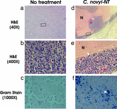
Fig. 1.
Tumor inflammation after i.v. injection with C. novyi-NT spores. Untreated tumors (a-c) and tumors treated with C. novyi-NT spores (d-f) were examined histopathologically after hematoxylin/eosin staining. Extensive areas of necrosis (N) were present within CT26 tumors 24 h after systemic injection of spores, and a ring of inflammation surrounded the tumors (e). The boxed areas in a and d are magnified in b and e, respectively, showing neutrophilic infiltrate in the treated lesions (e) but only tumor cells in the untreated control (b). The great majority of the cells in d and e were inflammatory cells (white arrow), with only a few nests of tumor cells remaining (red arrows). Gram stains (c and f) showed that the necrotic regions of only the treated lesions were filled with vegetative C. novyi-NT bacteria (white arrow).

