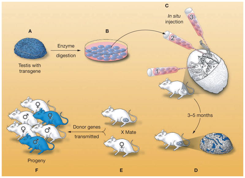
Figure 2.
Procedure for testis-cell transplantation as developed in the mouse. (A) A single-cell suspension is prepared from the testes of a fertile male that expresses a reporter transgene, Escherichia coli lacZ. (B) The testis cells can be cultured with appropriate conditions. (C) Cells are microinjected into the seminiferous tubules of an infertile recipient male. There are three methods for microinjection: the micropipette can be inserted (1) directly into the seminiferous tubules, (2) into the rete testis, or (3) into an efferent duct. (D) Spermatogonial stem cells colonize the basement membrane of the tubules and generate donor-cell-derived spermatogenesis, which can be stained blue using a substrate for the reporter gene product (β-galactosidase). Each blue stretch of cells in the seminiferous tubules of the recipient testis represents a spermatogenic colony derived from a single donor stem cell. (E) Mating the recipient male to a wild-type female results in donor-cell-derived spermatozoa fertilizing wild-type oocytes. (F) Progeny with the donor haplotype are produced. Modified with permission from references 19 © (2002) American Association for the Advancement of Science and 32 © (1997) University of the Basque Country Press.

