Figure 3.
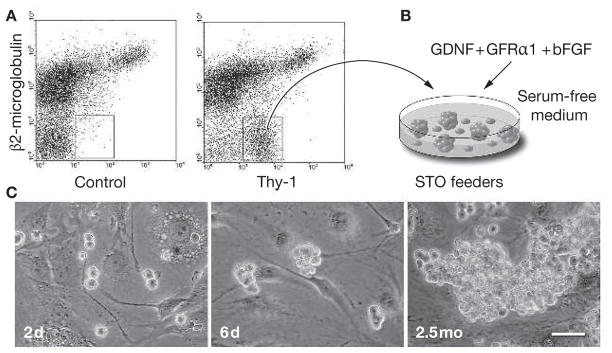
In vitro proliferation of mouse spermatogonial stem cells. (A) A highly enriched spermatogonial stem-cell population is obtained from adult cryptorchid mouse testes by fluorescence-activated cell sorting using antibodies against β2-microglobulin (the light chain of the MHC class I molecule) and Thy-1.51,52 The concentration of stem cells in the cryptorchid testes is 20-fold to 25-fold higher than in wild-type testes.76 (B) The enriched stem-cell population is placed on STO feeder cells in a serum-free defined medium supplemented with glial-cell-line-derived neurotrophic factor, soluble glial-cell-line-derived neurotrophic factor-family receptor α1 and basic fibroblast growth factor. The MHC class I− Thy-1+ cells form germ-cell clumps and proliferate. (C) Microscopic appearance of germ-cell clump formation and continuous proliferation of clump-forming cells is shown (2 days, 6 days and 2.5 months after in vitro culture of spermatogonial stem cells sorted using fluorescence-activated cell sorting; scale bar = 50 μm). The clump-forming germ cells can reconstitute normal spermatogenesis and restore fertility following transplantation into recipient testes of infertile male mice, indicating that they are spermatogonial stem cells. Under these culture conditions, spermatogonial stem cells continuously proliferate over 6 months.3 bFGF, basic fibroblast growth factor; d, days; GDNF, glial-cell-line-derived neurotrophic factor; GFRα1, glial-cell-line-derived neurotrophic factor-family receptor α1; mo, months.

