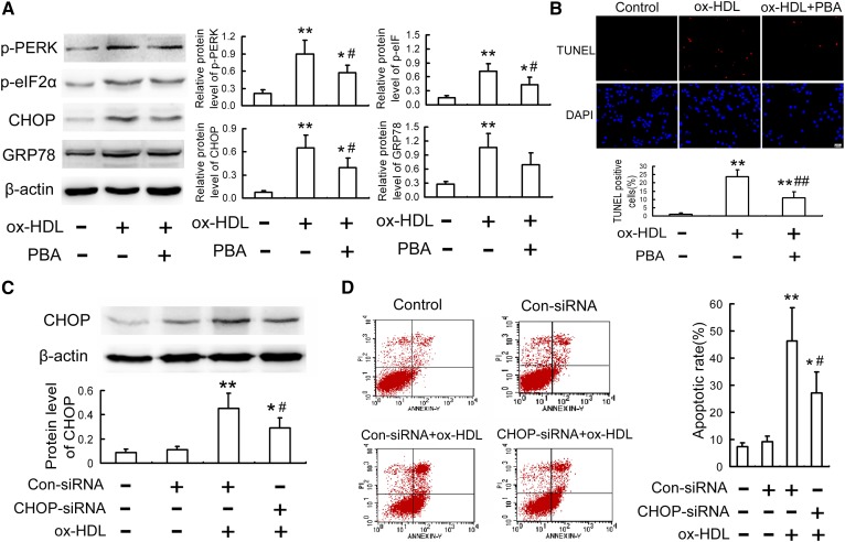
Fig. 3.
Alleviation of ER stress-CHOP pathway mitigates ox-HDL-induced macrophage apoptosis. A, B: RAW264.7 cells were treated with 100 mg/l ox-HDL in the presence or absence of PBA (5 mmol/l) for 24 h, and then the protein levels of ER stress markers and apoptosis were detected by Western blot and TUNEL assay, respectively. Scale bar = 20 µm. C, D: RAW264.7 cells were transfected with siRNA specific for CHOP, followed by treatment with 100 mgl ox-HDL for 24 h, and then CHOP level and apoptosis were determined by Western blot and flow cytometry, respectively. Data are expressed as the mean ± SD of at least three independent experiments. Con, control. *P < 0.05; **P < 0.01 versus control group; #P < 0.05; ##P < 0.01 versus ox-HDL group.

