Fig. 7.
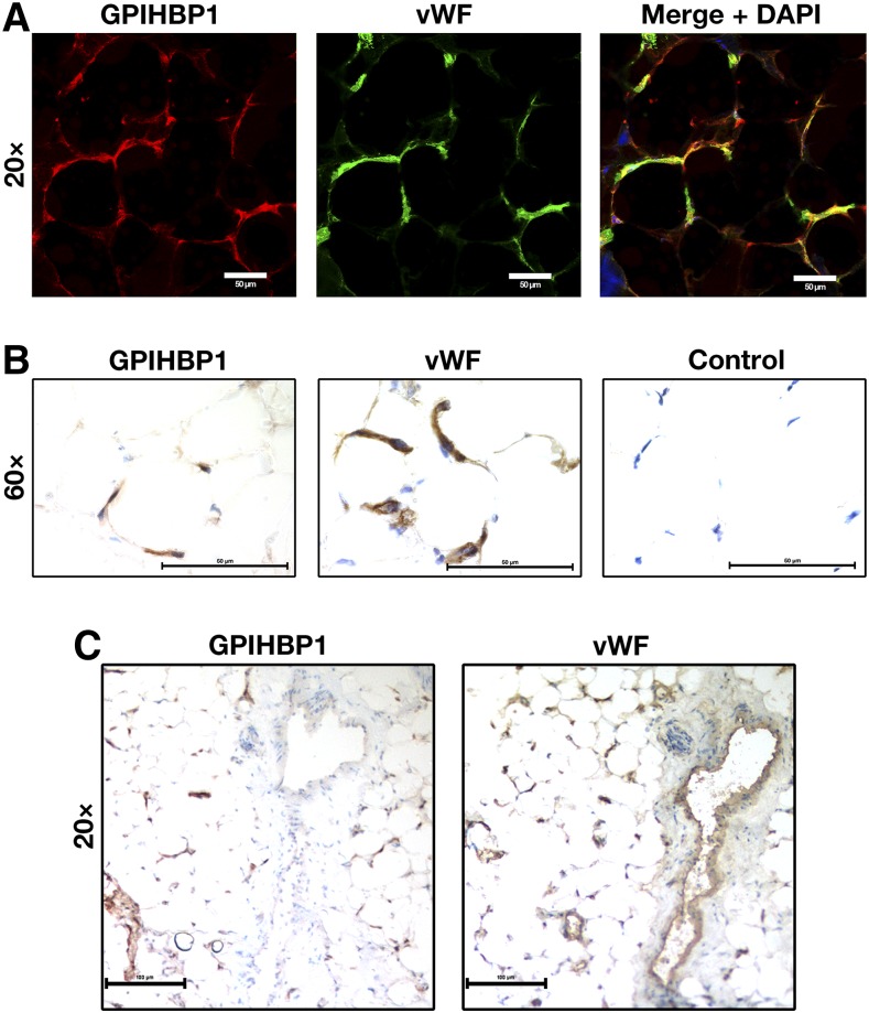
Detection of GPIHBP1 in human tissues with GPIHBP1-specific monoclonal antibodies. Sections of human cardiac adipose tissue (20 μm) were fixed with 3% paraformaldehyde and processed for confocal immunofluorescence microscopy (A) or light microscopy (B, C). (A) Confocal microscopy images showing GPIHBP1 (in this case, detected by a combination of mAbs RE3 and RF4, 10 μg/ml each; red) and von Willebrand Factor (vWF, a marker for endothelial cells; green) in the capillaries of human cardiac adipose tissue. (B) Consecutive HRP-stained sections showing GPIHBP1 (left panel, mAb RF4, 1 μg/ml) and vWF (middle panel) in capillaries of human cardiac adipose tissue. No primary antibody was added in the control panel (right). (C) Consecutive HRP-stained sections showing expression of vWF in both capillaries and a large venule, whereas GPIHBP1 was expressed in endothelial cells of capillaries but not in endothelial cells of the venule. In B and C, sections were counterstained with hematoxylin. Scale bar for A, 20 μm; B, 50 μm; C, 100 μm.

