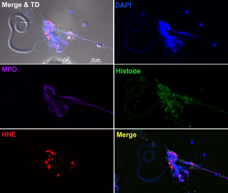
Fig 1. B. malayi Mf entangled within neutrophil extracellular traps.
Fluorescence imaging of a worm-NET aggregate labelled with DAPI (blue) and antibodies to myeloperoxidase (MPO; purple), citrullinated histone H4 (histone; green) and human neutrophil elastase (HNE; red). The co-localization of markers with Mf is shown in the merged image with transmitted light (top left panel) and the overlap of MPO, histone and HNE with DNA shown in the merged image (bottom right panel). Live Mf were incubated in the presence of human neutrophils for 18 hours at 37°C and 5% CO2. Scale bar = 10μm.

