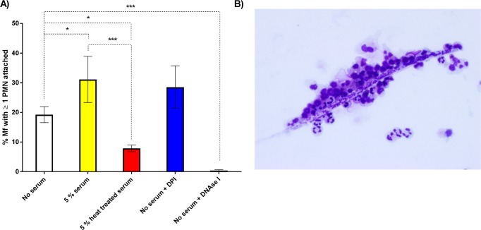
Fig 3. The attachment of PMNs to B. malayi Mf.
A: The percentage of Mf that had at least one PMN adhered to their surface or indirectly fastened by extracellular DNA at 24 hours post-experimental set up (n≥6). White, yellow, red, blue, and black bars represent no serum, 5% autologous serum, 5% autologous heat-treated (55°C, 30 mins) serum, 10μM diphenyleneiodonium (DPI) and 30μg/ml DNase I treated wells respectively. Error bars represent standard error of the mean; *P<0.05, **P<0.01, ***P<0.001. B: Image of PMNs adhered to a Mf. Image taken after 24 hours of incubation in 5% autologous serum at 37°C and 5% CO2. The preparation was stained with modified Wrights’ stain.

