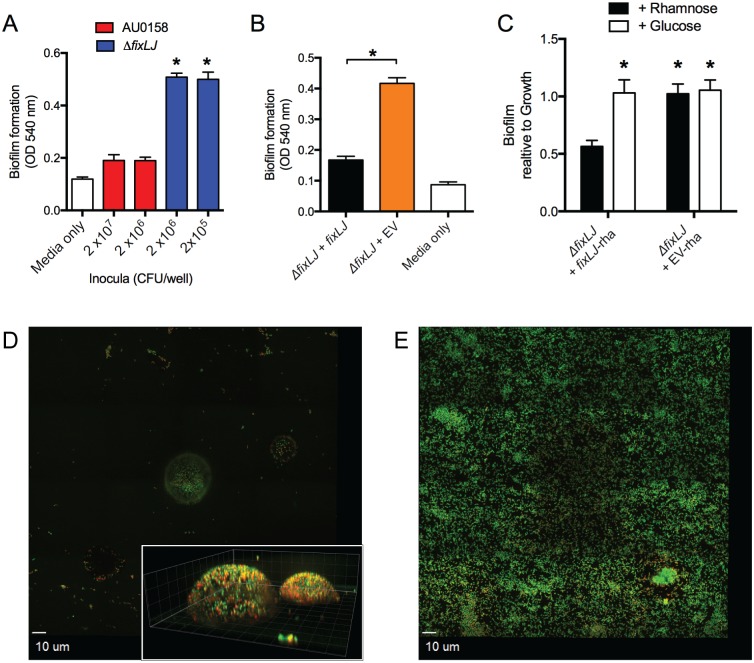
Fig 4. The B. dolosa fixLJ deletion mutant produces more biofilm by crystal violet staining and has a different biofilm structure.
Biofilm formation of B. dolosa AU0158 constructs on PVC plates as measured by crystal violet staining at 48 hours. (A) B. dolosa strain AU0158 produces less biofilm than its fixLJ deletion mutant. Strains were grown in TSB with 1% glucose at varying inocula. (B) The B. dolosa fixLJ deletion mutant complemented with fixLJ under the control of its own promoter produces less biofilm compared to the strain carrying an empty vector (EV). (C) A B. dolosa fixLJ deletion mutant complemented with fixLJ under the control of a rhamnose-inducible promoter or empty vector grown in LB in the presence of glucose (0.4%, which represses the promoter) or rhamnose (0.4%) and compared to the ΔfixLJ + fixLJ strain grown in rhamnose-containing medium. For panels A-C, bars represent mean measurements of 5–6 replicates and error bars represent one standard deviation (representative of three independent experiments). *P<0.05 compared to AU0158 by 1-way ANOVA with Tukey’s multiple comparison test. Representative mosaic images of biofilms of strain AU0158 (D) and its fixLJ deletion mutant (E) grown on 8-well chamber slides for 48 hours, stained with live/dead stain, and imaged by confocal microscopy. For panels D and E, live bacteria are stained green and dead are red. The inset of panel D shows Z-stack images taken at 1 μm intervals; gridlines denote 5 μm lengths. Images are representative of two independent experiments conducted with four replicates.

