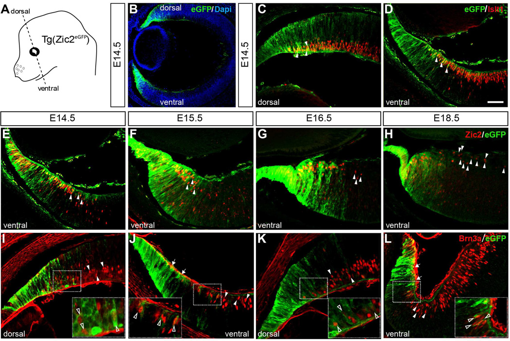
Figure 1. Spatiotemporal expression of eGFP in the CMZ and peripheral neural retina of Tg(Zic2eGFP) embryos.
A. Scheme of an embryonic mouse head showing the orientation of coronal sections.
B. Retinal section of a E14.5 Tg(Zic2eGFP) embryo showing eGFP staining in dorsal and ventral peripheral retina.
C–D. E14.5 Tg(Zic2eGFP) embryo stained for Islet1 reveals that some RGCs in the peripheral retina express eGFP in both the dorsal and the ventral retina (white arrowheads).
E–H. Zic2 staining in ventral retinal sections of Tg(Zic2eGFP) embryos at the indicated stages show that eGFP colocalizes with Zic2 in the CMZ and peripheral neural retina but that many Zic2+ cells within the peripheral neural retina are not eGFP positive (white arrowheads).
I–L. Brn3a staining in retinal sections of Tg(Zic2eGFP) embryos at the indicated stages shows that some Brn3a+ cells are also eGFP+ in both dorsal and ventral retina (open arrowheads), while some Brn3a+ are eGFP− (white arrowheads). Note the gap of Brn3a+ cells in the ventral retina (white arrows in panels J and L).
Scale bar 50 µm.

