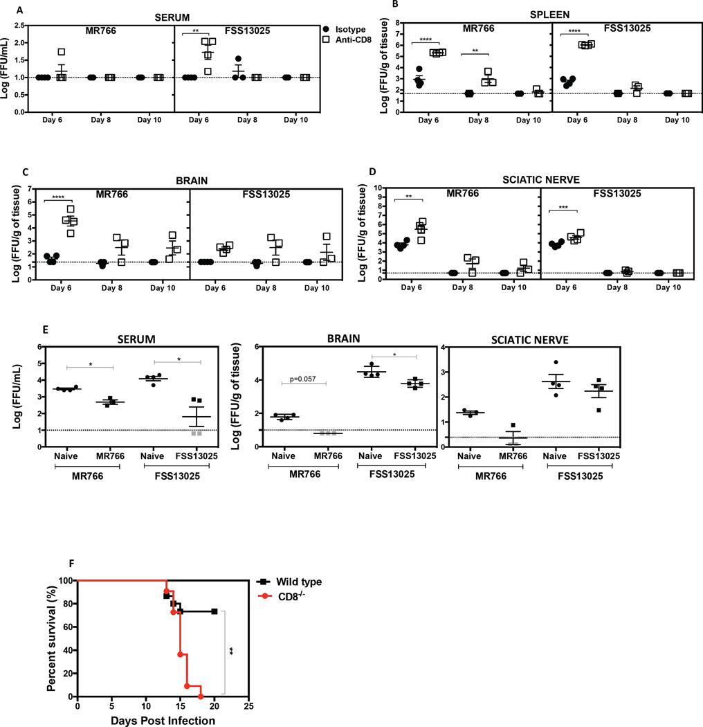
Figure 6. Protective role of CD8+ T cells against ZIKV infection in mice.
LysMCre+IFNARfl/fl were treated with depleting anti-CD8 or isotype control antibody on days 3 and 1 before infection with 105 FFU of MR766 or FSS13025. Mice were sacrificed and tissues harvested at 6, 8 and 10 days post-infection. The levels of infectious virus in the (A) serum, (B) spleen, (C) brain, and (D) sciatic nerve were quantified using BHK-21 cell-based FFA. A two-way ANOVA test was used to compare the levels of infectious ZIKV between the isotype and the anti-CD8 antibody-administered groups for all time points and tissues. (E) On day 120 after infection with MR766 or FSS13025, 7.5 × 10⁶ CD8+ T cells were transferred into 5-week-old naive mice one day before challenge with 105 FFU of MR766 or FSS13025. For controls, CD8+ T cells were isolated from naive LysMCre+IFNARfl/fl mice. Infectious ZIKV was quantified. Mann-Whitney test was used to compare naïve CD8+ T cells vs. ZIKV-immune CD8+ T cells. (F) Seven-week-old WT and CD8α−/− were treated with 2 mg of IFNAR-blocking antibody at day -1, and then inoculated subcutaneously with 105 PFU of mouse adapted Dakar 41519 ZIKV strain at day 0. Survival was monitored for 21 days in both groups and reported for WT (n=15, Black square) and CD8−/− (n=11, Red circle) mice. Pooled data from three independent experiments are represented and the log-rank (Mantel-cox) test was used to compare groups.

