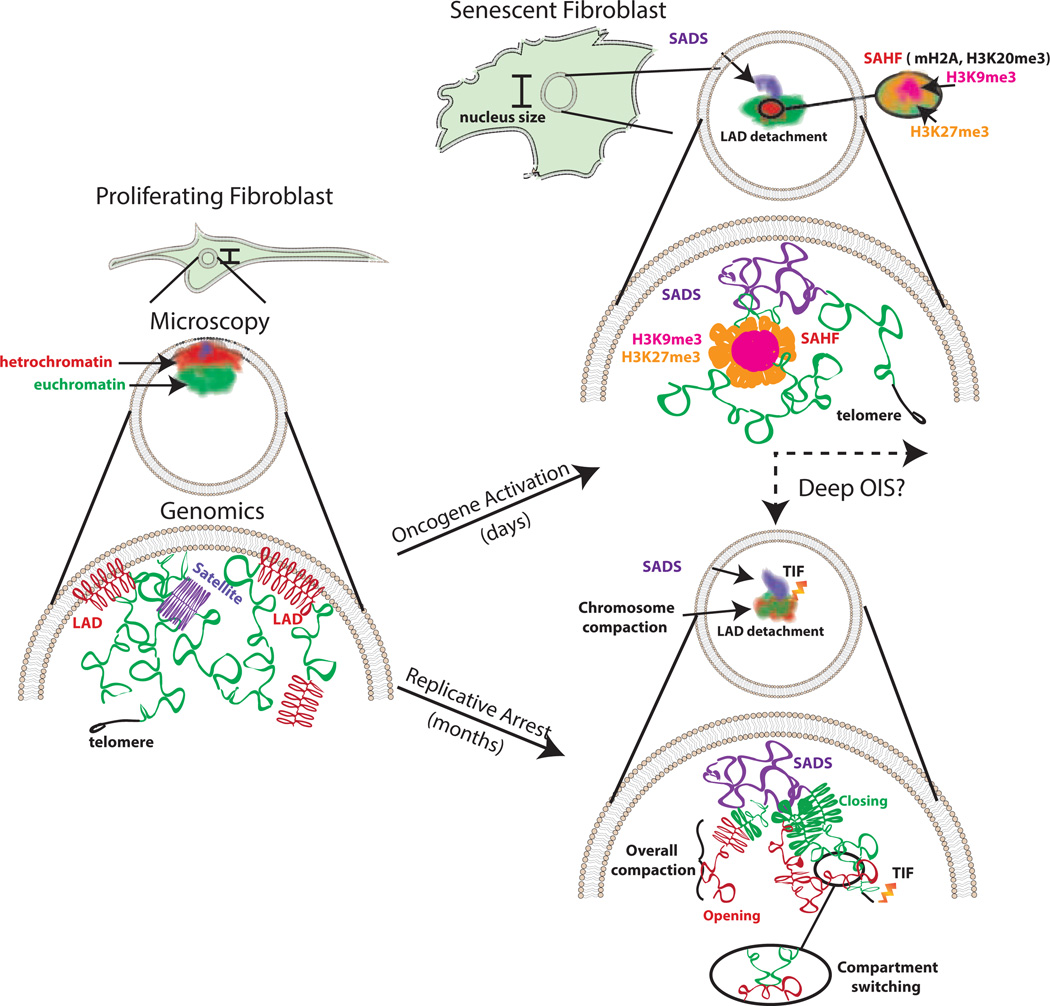
Figure 1. Key Figure: Changes in chromosome organization in OIS and RS.
Summary of the alterations to chromatin that occur in senescent cells observed by microscopy and genomics techniques. In proliferating cells, heterochromatin and the centromere tend to associate with the nuclear periphery and the euchromatic regions are found in the nuclear interior. The centromere is densely packed. Euchromatin is accessible for transcription. Peripheral heterochromatin is tethered to the nuclear lamina in regions called LADs. In cellular senescence, conserved and distinct features are observed depending on the stimuli. Shared features include SADS formation and the loss of interaction with the nuclear periphery or LADs detachment. SAHF appear to be predominantly induced in OIS and show foci with an H3K9me3 interior and an H3K27me3 halo. Other features, such as chromosome shrinkage, focal hypermethylation/decreased accessibility of euchromatic regions, hypomethylation/increased accessibility of constitutive heterochromatin and compartment switching, have only been investigated in RS but might be present in other types of senescence as well. Telomere Dysfunction-Induced Foci (TIF) are prominent in RS [98–100]; however, some evidence suggests this feature may be present in OIS [101].

