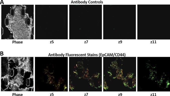
Fig 3. Evaluation of antibody perfusion through the tumor FNAB on device.
(A) 10x Phase contrast image of FNAB sample in trap of device and 10x fluorescent z-axis images (z5, z7, z9, z11) 24 hours post the staining procedure using isotype control antibodies for both EpCAM (red-Cy5) and CD44 (green-FITC). (B) 10x Phase contrast image of FNAB sample in trap of device and 10x fluorescent z-axis images (z5, z7, z9, z11) 24 hours post the staining procedure using EpCAM (red-Cy5) and CD44 (green-FITC).

