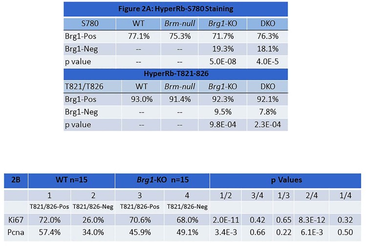
Figure 2. Figure 2A shows the results of dual IF staining with anti-Brg1 and anti-pRb1S780 (top) or anti-pRb1T821/826 (bottom) in lung tumors derived from each of the four genotypes.
The results from Brg1-positive tumor cells (top row) are compared with results from Brg1-negative tumor cells, and the p values (bottom rows) from the comparisons are given. Figure 2B shows the results for dual IF of pRb1T821/826 with anti-Ki67 (top row) and with Pcna (bottom row). P values that compare the percentage of positive staining of Ki67 and Pcna when pRb1T821/826 staining is either positive or negative are given.

