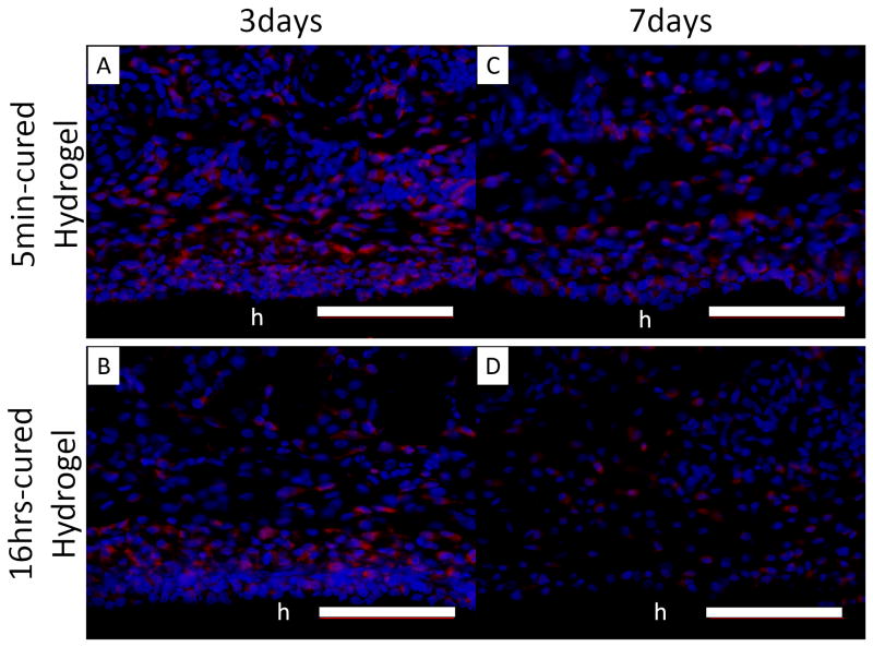
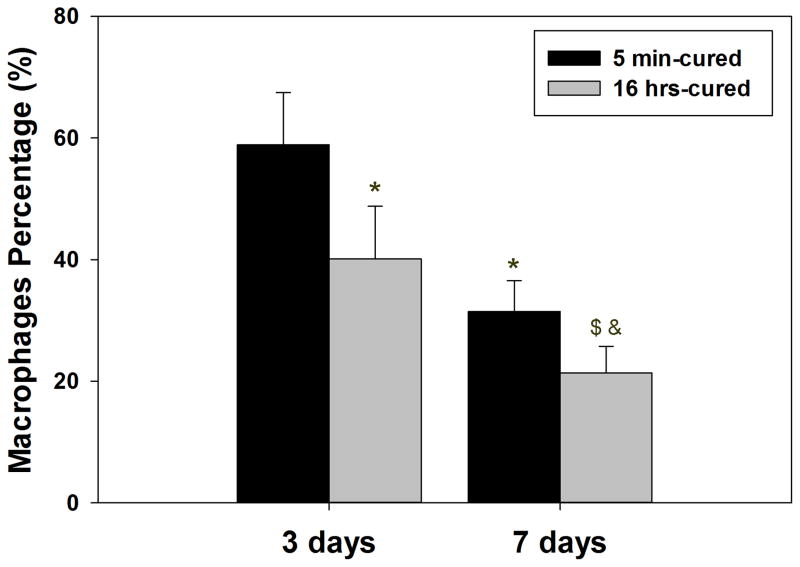
Figure 9.
Immunofluorescent staining of macrophages (CD 68) in tissue surrounding 5 min- and 16 hrs-cured PEG-D4 hydrogels after 3 (A, B) and 7 (C, D) days of subcutaneous implantation. Cell nuclei and macrophages were stained by DAPI (blue) and CD68 (red), respectively. The letter h indicates the location of the implanted hydrogel. The percentage of macrophages relative to the total number of cells stained with DAPI (E). * p < 0.05 when compared to 5 min-cured hydrogels after 3 days of subcutaneous implantation; $ p < 0.05 when compared to 5 min-cured hydrogels after 7 days of subcutaneous implantation; & p < 0.05 when compared to 16 hrs-cured hydrogels after 3 days of subcutaneous implantation. Scale bar: 100 μm.


