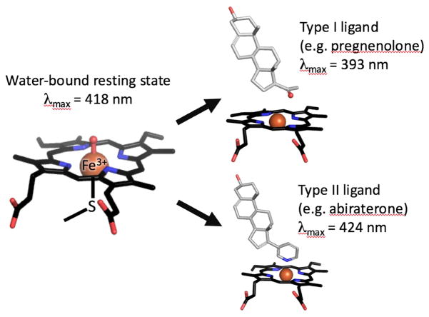
Figure 2.
Heme-iron in different coordination state is shown by stick model. Absorbance maxima at 417 nm is representative of water coordinated oxidized heme-iron (resting state), while type I ligand and type II ligand shift absorbance maxima towards blue spectrum (393 nm) and red spectrum (424 nm), respectively.

