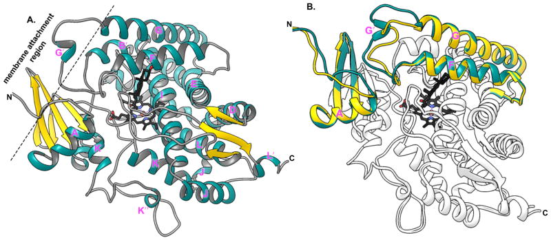
Figure 3.
Ribbon representation of crystal structure of CYP17A1 highlighting characteristic structural features. A. Structure of progesterone (black sticks) bound CYP17A1 A105L mutant (PDB id: 4nkx) showing typical P450 structural folds with iron coordinated heme in the active site. B. Overlay of the two molecules of CYP17A1 A105L from one crystal symmetric unit is shown. A/B (shown in dark cyan) and C/D (gold) highlight the variation in the backbone structure of helix F and G structure, which interacts with the ER membrane.

