
Figure 3. Proteasomal-Dependent Outer Mitochondrial Membrane Rupture Is Required for PHB2/LC3 Interaction and Mitophagy.
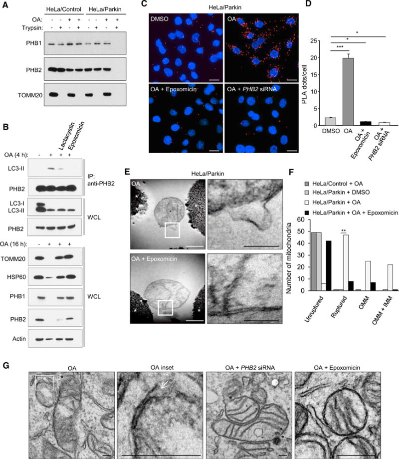
(A) Protease protection assay of mitochondrial fractions purified from HeLa/Control or HeLa/Parkin cells treated with DMSO or OA for 4 hr.
(B) Effects of proteasome inhibitors (5 mM lactacystin or 100 nM epoxomicin) on PHB2/LC3-II interaction, OMM rupture and mitophagy in HeLa/Parkin cells treated with OA for 4 hr (top gels assess co-immunoprecipitation of PHB2 and LC3) or 16 hr (bottom gels show western blots of indicated mitochondrial proteins). WCL, whole cell lysates. Similar results were observed in two independent experiments.
(C and D) Representative images (C) and quantification (D) of Duolink in situ PLA assay demonstrating the interaction between LC3 and PHB2 in HeLa/Parkin/Flag.StrepII-LC3 cells in cells transfected with NC or PHB2 siRNA for 48 hr and then treated for 2 hr with either DMSO, OA, or OA + epoxomicin. In (D), bars are mean ± SEM for triplicate samples of >50 cells per sample. Similar results were obtained in three independent experiments. ***p < 0.001, *p < 0.05;one-way ANOVA.
(E) Electron micrographs of mitochondria immunoprecipitated from HeLa/Parkin cells treated for 4 hr with OA in the presence or absence of epoxomicin. Right panels: higher magnification images of regions outlined in left panels. See also Figure S2. Asterisk, Dynabead. White scale bar, 500 nm. Black scale bar, 200 nm.
(F) Quantitation of mitochondria with unruptured or ruptured membranes (further subdivided into mitochondria with OMM rupture only or rupture of both OMM and IMM [OMM + IMM]) in the experiment shown in (E). Fifty mitochondria per condition were examined by an observer blinded to experimental condition. **p < 0.01, Chi-square test.
(G) Representative electron micrographs showing mitochondrial rupture in HeLa/Parkin cells transfected with NC or PHB2 siRNA and treated with OA with or without epoxomicin for 2 hr. White asterisk and white arrow, site of focal OMM rupture; black asterisks, sites of focal rupture of OMM and IMM. Scales bars, 100 μm.