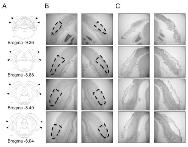
Figure 2. POR lesions.
A) The rostrocaudal extent of the postrhinal cortex shown in coronal sections on a line drawing. B–C) photomicrographs taken from a rat with no lesion (B) and from one animal with an extensive POR lesion (C). Damage ranged from 75–98% bilaterally across the entire rostrocaudal extent of the POR in 9 animals (mean damage = 88 ± 2%). The brain depicted in C had 85% damage to the POR. Arrowheads (A) and dashed lines (B) demarcate the borders of the POR.

