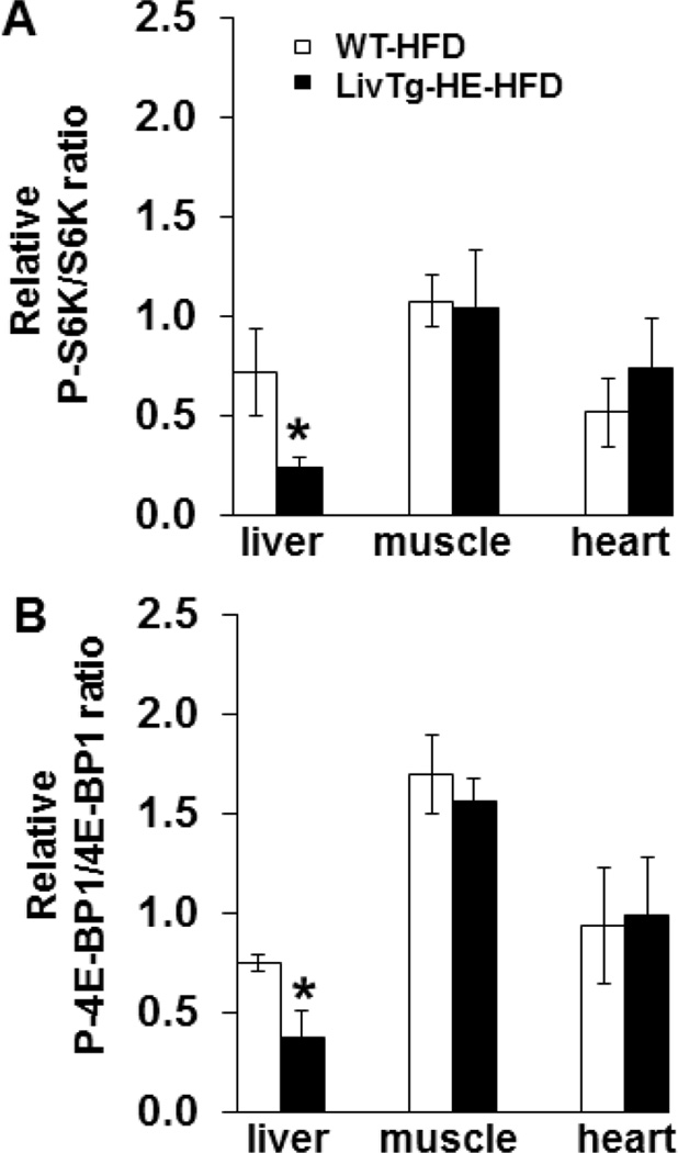
Fig. 7.
Phosphorylation of S6K and 4E-BP1 is lower in the liver of LivTg-HE mice fed a high fat diet (HFD) compared to WT mice fed the same diet. P-S6K, S6K, P-4E-BP1, and 4E-BP1 were determined by Western Blotting in liver, muscle, and heart of Leu-gavaged WT-HFD and LivTg-HE-HFD mice. Animals were sacrificed 30 min after Leu gavage. Each graph shows the relative ratio between the phosphorylated and total form of S6K (A) and 4E-BP1(B). Data represent mean ± SEM, n=5–6 male mice, age 24 weeks. *P≤ 0.05 as compared to WT-HFD.

