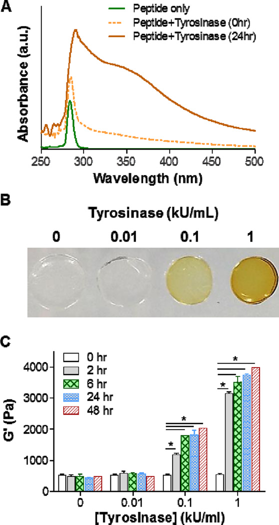
Figure 2. Tyrosinase-mediated in situ stiffening of PEG-based hydrogels.
(A) UV/Vis absorbance spectrum of 5 mM CYGGGYC peptide, peptide/tyrosinase (1 kU/mL) mixture before (0hr) and after 24hr incubation. (B) Photographs of PEG-peptide (i.e., 2.5wt% PEG8NB, 5 mM CYGGGYC) hydrogels treated with different concentrations of tyrosinase. (C) Effect of tyrosinase concentration of shear moduli (G’) of the PEG-peptide hydrogels. Crosslinked hydrogels were incubated in PBS for one day prior to 6hr of tyrosinase treatment. Afterward, the gels were transferred to PBS and gel moduli were monitored periodically using oscillatory rheometer. Data represent Mean ± SEM (n = 3). Asterisks indicate p<0.05 (compared with gels at 0hr, i.e., prior to tyrosinase treatment).

