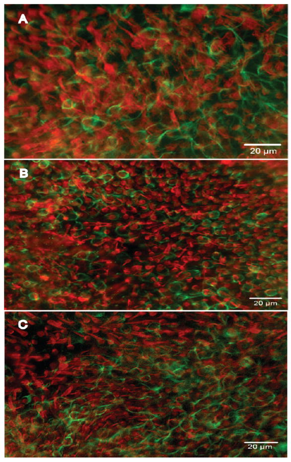
Figure 2.

Whole-mounts of utricles stained with antibodies to myosin VIIa (red) and neurofilament (green) viewed with epi-fluorescence. A. A normal (control) utricle displaying a confluent population of HCs. Rings of neurofilament around HCs denote a calyx-type nerve endings surrounding type I HCs. B-C. Utricles from animals treated with cadmium (B) and lead (C) in which the density of HCs and calyx-type nerve endings is similar to control.
