
Figure 2.
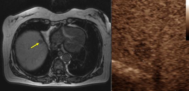
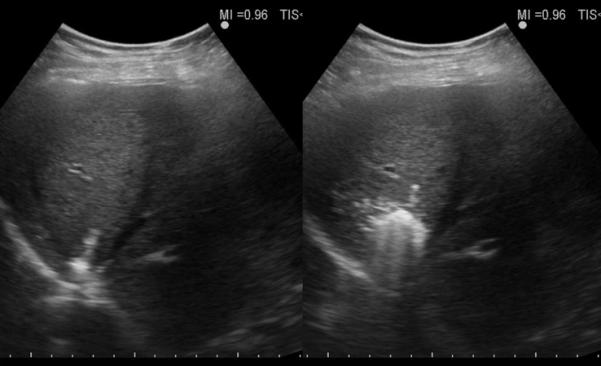
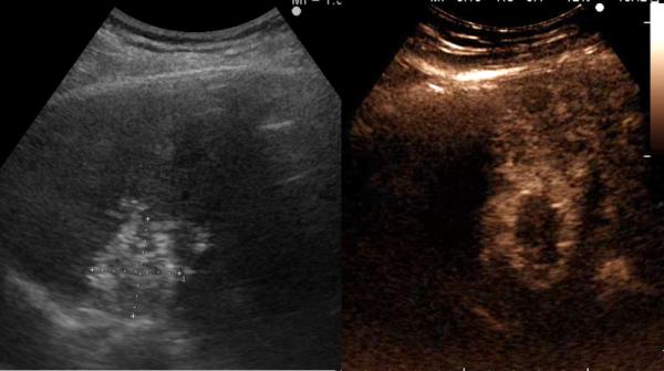
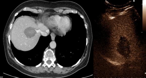
67-year old female with liver metastasis from colorectal cancer. A) Pre-ablation magnetic resonance (MR) imaging (left) of liver lesion (arrow) and CEUS (right) confirming presence of 13 by 14 mm liver lesion (arrow) B) The lesion was treated with a single water-cooled antenna at 50 W for 6 minutes (left). Post-ablation image demonstrating hyper-echoic region measuring 34 mm by 37 mm, representing the ablation zone (right). The goal is to create ablation margins > 1 cm beyond tumor boundary. C) Post-ablation B-mode ultrasound (left) and CEUS (right) showing the ablation zone (anechoic) with the presence of the hypervascular peri-lesional halo. D) CECT (left) and CEUS (right) demonstrating ablation zones that encompass the entire lesion measuring 32 by 42 mm.