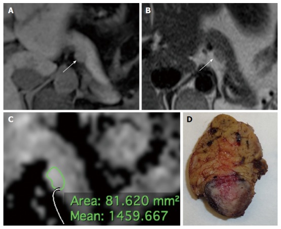
Figure 1.

Small hyperfunctioning pancreatic neuroendocrine neoplasm (insulinoma) classified as G1, stage 1 tumor at histology (Ki67 < 1%, T1, N0, M0) in a 44 years-old woman. A: Axial volumetric interpolated breath-hold examination (VIBE) gradient echo image (repetition time msec/echo time msec, 4.3/1.4) with fat suppression shows a homogeneously hypointense lesion with well defined margins (arrow) in the pancreatic body; B: On axial T2-weighted half-Fourier single-shot turbo spin echo (HASTE) image (TR/TE, ∞/90), the tumor appears slightly hyperintense (arrow) compared to adjacent pancreatic parenchyma; C: At ADC quantification the tumor had a relatively high mean ADC value; D: The macroscopic pathological specimen (distal pancreatectomy, sagittal cut) shows a small, well-delimitated lesion bulging the contour of the pancreatic body.
