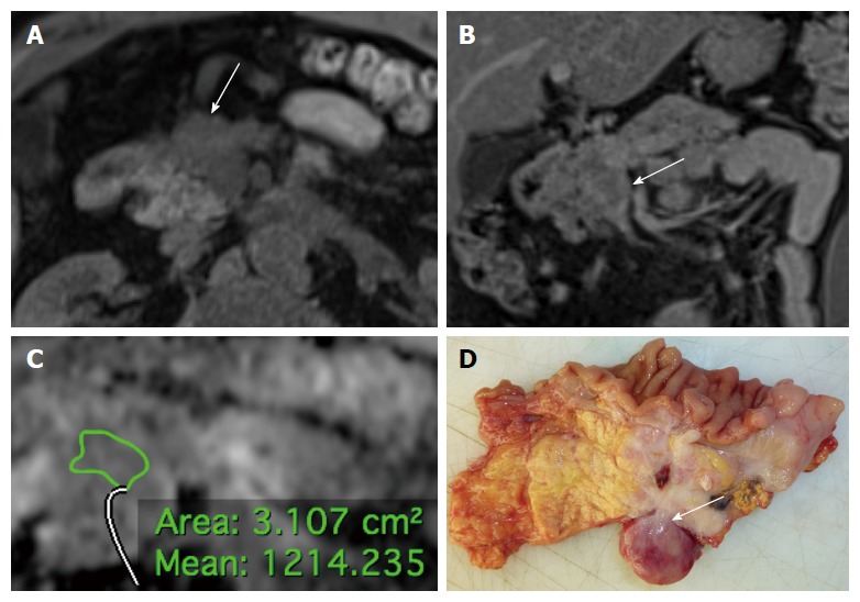
Figure 2.

Non-hyperfunctioning pancreatic neuroendocrine neoplasm of the pancreatic head classified as G2, stage 3b tumor at histology (Ki67 15%, T4, N1, M0) in a 55 years-old man. A: Axial volumetric interpolated breath-hold examination (VIBE) gradient echo image (repetition time msec/echo time msec, 4.3/1.4) with fat suppression shows a hypointense tumor with ill-defined margins (arrow); B: On coronal fat-saturated T1-weighted volumetric interpolated breath-hold examination (VIBE) gradient echo image (TR/TE 3.5/1.3 ms) acquired during the delayed phase of the dynamic study the tumor shows heterogeneous contrast enhancement, with infiltration of the superior mesenteric vein (arrow); C: The tumor presents intermediate mean ADC value; D: The surgical specimen (pancreaticoduodenectomy with en bloc resection of the superior mesenteric vein, transverse cut) shows a large lesion of the pancreatic head with irregular margins infiltrating the superior mesenteric vein (arrow).
