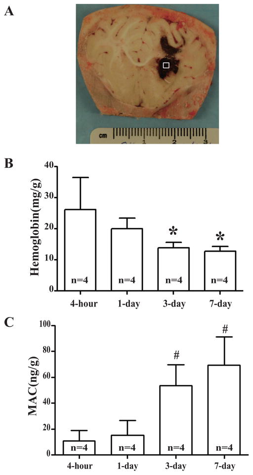
Figure 2.
(A) Coronal section of an in situ frozen piglet brain after ICH. The square represents the sample area. (B) Time course of hemoglobin content in hematoma. Values are mean ± SD, *p <0.05, #p <0.01 vs. 4-hour. (C) Time course of MAC content in the hematoma. Values are mean ± SD, *p <0.05, #p <0.01 vs. 4-hour.

