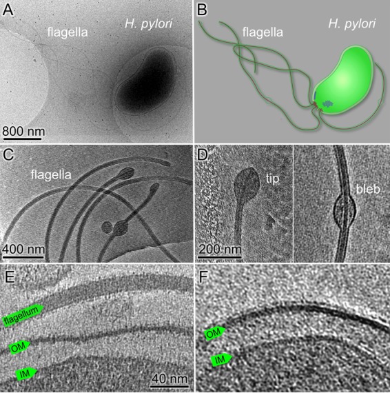
FIG 1.

Ultrastructure of H. pylori cells revealed by cryo-ET. (A) An overall cryo-EM image of a wild-type H. pylori cell with many flagella. (B) A schematic model of the H. pylori cell with particular emphasis on the cell envelope, flagella, and chemoreceptor arrays. (C) A tomographic slice shows the sheathed flagella with membrane blebs at the tip or in the middle of the flagella. (D) Zoomed-in views of a membrane bulb at the flagellar tip (left) and a membrane bleb in the middle of a flagellum (right). (E) A tomographic slice at the cell pole shows a sheathed flagellum and the cell envelope, including the outer membrane (OM) and the inner membrane (IM). (F) Fine filamentous features that cover the outer membrane and the flagellar sheath presumably represent bacterial lipopolysaccharide (LPS).
