FIG 3.
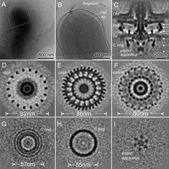
Flagellar motor structure of H. pylori revealed by cryo-ET and subtomogram averaging. (A) A typical cryo-EM image of one bacterium at low magnification. (B) A central slice of the tomogram from the cell pole outlined in panel A. Note that one flagellar motor is embedded in the cell envelope, including the outer membrane (OM) and the inner membrane (IM). (C) A central slice of the averaged structure. (D to F) Three cross sections of the flagellar motors show distinct features with 18-fold symmetry in periplasmic space. (G and H) Two cross sections from the top and the bottom of the C ring, respectively. (I) One section from the export apparatus shows a hexagonal pattern.

