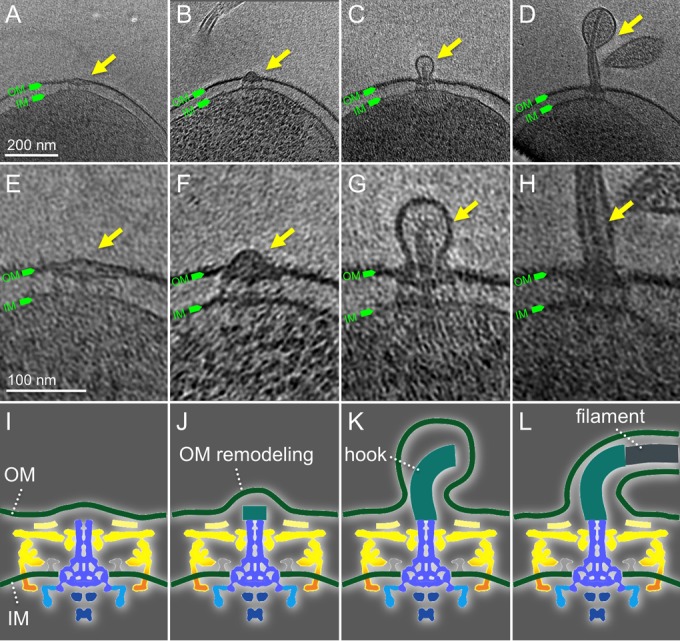
FIG 6.

The assembly of flagella couples with the biosynthesis of the outer membrane sheath. (A to D) Representative slices of cryo-ET reconstructions show different intermediates during flagellar assembly. (E to H) The corresponding zoomed-in views of images from panels A through D. The corresponding cartoon illustrations show different stages: rod assembly (I), hook assembly (J, K), and filament assembly (L). The following color scheme is the same as that in Fig. 4: yellow, cage; orange, stator; green, membranes; blue, rod and MS ring; cyan, C ring; and dark blue, export apparatus.
