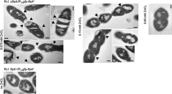
FIG 4.
Cell division septa are abnormal in filamentous cells after moderate FtsA depletion. Representative micrographs of ultrathin cell sections showing morphological defects of cells depleted of FtsA in TSB at 37°C. Samples for microscopy were taken 4 h after the start of FtsA depletion (at the indicated levels of ZnCl2) and washed, fixed, and vitrified, as described in Materials and Methods. Septation defects characteristic of each depletion condition are indicated by arrows. Scale bars, 0.2 μm.

