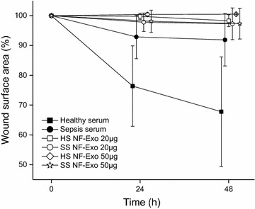
Fig. 4.

Keratinocyte migration in wounded monolayer after exposure to exosomes. Exosomes derived from healthy sera (HS NF-Exo) and sepsis sera (SS NF-Exo) treated normal fibroblasts were used in 20 or 50 µg/ml concentrations. Wound area (%) reduction was followed 48 h and data are presented as means with standard deviations from seven wounds per group
