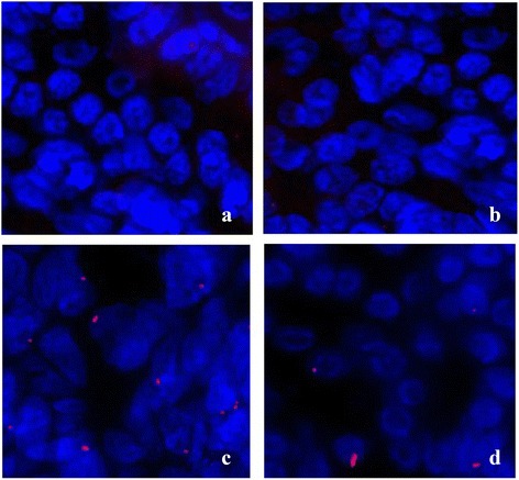
Fig. 10.

Immuno cytochemistry of Eotaxin in groups. Eotaxin expression in control, AR-NS, AR-HRS and Normal-HRS groups was determined by immuno cytochemistry. Immunoreactive eotaxin, detected using a Cy3-labeled secondary antibody (red), was highly expressed in nasal mucosa of AR-NS group (c), and decreased in AR-HRS group (d). While in control subjects and normal-HRS subjects eosinophil infiltration was very scarce (a and b)
