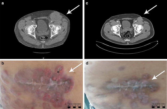
Fig. 1.

Computed tomography (CT) and macroscopic images of the inguinal lesion before and after 3 cycles of chemotherapy. a CT scan shows a subcutaneous metastatic melanoma lesion (arrow) of 76 mm × 63 mm in the left inguinal area before chemotherapy. b Cutaneous metastatic melanoma lesions (arrow) were nodular and inflammatory before chemotherapy. c CT scan shows that the size of the subcutaneous metastatic melanoma lesion (arrow) decreased to 31 mm × 35 mm, with a reduction of 48%, after 3 cycles of chemotherapy. d Cutaneous metastatic melanoma lesions (arrow) exhibited massive shrinkage, leaving a fibrotic quality of the skin, after 3 cycles of chemotherapy
