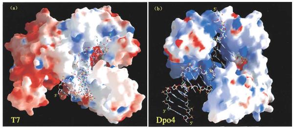
Fig. 3.
Crystal structures of T7 DNA polymerase and Dpo4 containing DNA. (a) T7 DNA polymerase and (b) Dpo4 are shown in molecular surface representation with positive (blue) and negative (red) electrostatic potential. The DNA molecules are drawn in sticks. The minor groove adjacent to the replicating base pair is exposed to solvent for Dpo4 but completely buried for T7 DNA polymerase. These figures are from Ling et al. Cell107 (2001), 91-102 [40].Copyright © 2001 Cell Press. All rights reserved.

