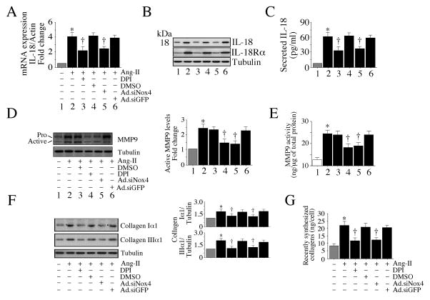
Fig. 3. Ang-II induces IL-18 and MMP9 expression via Nox4 and ROS.
A, B, C, Ang-II induces IL-18 and IL-18Rα expression and IL-18 secretion. CF infected with Ad.siNox4 (MOI 100 for 24 h) or treated with DPI (10 μM for 30 min) prior to Ang-II (10−7M) treatment for 2 (A, B) or 24 h (C) were analyzed for IL-18 mRNA expression by RT-qPCR (A, n=6), mature IL-18 (18 kDa form) and IL-18Rα protein levels in cleared whole cell lysates by immunoblotting (n=3), and secreted IL-18 levels in equal amounts of culture supernatants by ELISA (n=6). D, E, Ang-II induces MMP9 activation. The CF made quiescent in RPMI 1640 medium supplemented with ITS were treated with Ang-II (10−7M) for 2 (D) or 24 h (E). The culture supernatant was concentrated and 1μg/sample was analyzed by immunoblotting using antibodies that detect both pro and active forms of MMP9 (D). Densitometric analysis of the immunoreactibe bands from three independent experiments is summarized on the right. Enzymatic activity was also analyzed by a Biotrak activity assay kit (E). E, *P < 0.01 vs. respective untreated; †P < at least 0.05 vs. Ang-II ± Ad.siGFP or DMSO (n=6). F, G. Ang-II increases collagen expression. The quiescent CF were treated with Ang-II (10−7M) for 2 (F) or 72 h (G), and analyzed for collagens I and III by immunoblotting (F; n=3). Levels of soluble collagens released into culture media at 72 h were determined by Sircol™ collagen assay (G). A, C, *P < 0.001 vs. untreated. A–G, *P < at least 0.05 vs. respective untreated, †P < at least 0.05 vs. Ang-II (n=3–6).

