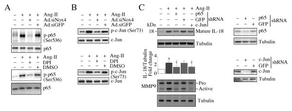
Fig. 4. Ang-II stimulates IL-18 and MMP9 expression via Nox4/ROS-dependent NF-κB and AP-1 activation.
A, Ang-II stimulates NF-κB/p65 activation via Nox4 and ROS. The quiescent CF were treated with DPI (10 μM in DMSO for 30 min) prior to Ang-II (10−7M for 30 min) addition. Phospho-p65 levels were analyzed by immunoblotting using cleared whole cell homogenates and activation-specific antibodies (upper and lower panels; n=3). B, Ang-II stimulates AP-1/c-Jun activation via Nox4 and ROS. The quiescent CF treated as in A were analyzed for phospho-c-Jun levels by immunoblotting using cleared whole cell homogenates and activation-specific antibodies (upper and lower panels; n=3). C, Ang-II induced IL-18 expression and MMP9 activation via NF-κB and AP-1. CF were infected with lentiviral p65 or c-Jun shRNA (MOI0.5) for 48 h. In the last 24 h, the complete media was replace with 0.5% BSA (IL-18) or RPMI 1640/ITS medium (MMP9), and then treated with Ang-II for 2 (IL-18) or 24 h (MMP9). IL-18 expression was analyzed by immunoblotting (n=3) using cleared whole cell lysates and antibodies that specifically detect the mature form (upper panel). MMP9 activity was analyzed using 1 μg of protein concentrated from culture supernatants (lower panel; n=3). Knockdown of p65 and c-Jun was confirmed by immunoblotting (right panels; n=3).

