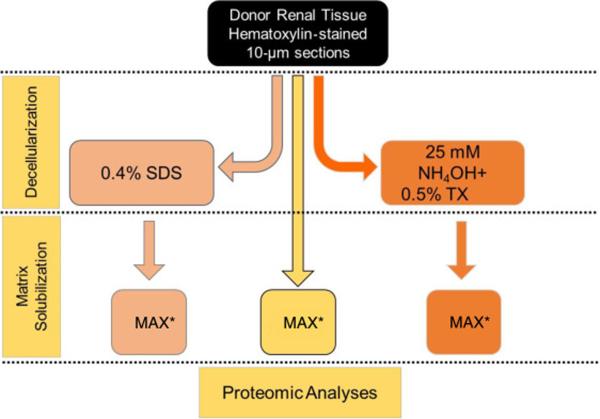
Figure 8. Optimization of ECM isolation for proteomic analyses.
A schematic workflow for three protein extraction methods comparison. Method (A) is a single step extraction/solubilization of tissue using the acid labile detergent Protease MAX surfactant with heating for 5min at 95ºC (MAX). Method (B) is the two step sequential extraction first with phosphate buffered saline 0.4% SDS to isolate the cellular fraction followed by ECM solubilization by MAX. Method (C) is the two step sequential extraction first with 25 mM NH4OH/0.5% TritonX-100 to isolate the cellular fraction followed by ECM solubilization by MAX.

