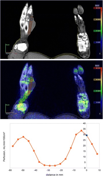
Fig. 3.

Abscess at metatarsus II in pig no. 4. Top: CT image with chosen VOI position reported in Table 4, determined from CT with guidance from [15O]water PET image. Middle: Fused PET/CT image with a profile of VOIs at vertical spacing of 2 voxels. Bottom: Perfusion profile from these VOIs. The chosen VOI corresponds to distance 0 mm and turns out not to be maximum value, but judged from other side of the profile, the value appears to be typical for the non-necrotic part of the abscess
