Fig. 1.

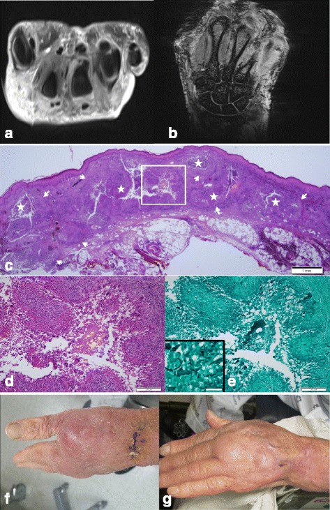
a MRI T2 weighted contrast transverse image reveals diffuse edematous swelling in the dorsum of the wrist, fluid in the extensor tendon sheath and fluid collection in the distal radio-ulnar joint. b MRI T1 weighted coronal image reveals diffuse paratendinous fluid in the extensor tendon sheath. c Skin biopsy shows multiple variable-sized granulomas (arrows) bearing central microabscesses (asterisks) in the dermis. H&E staining, Scale bar measures 1 mm. d Higher magnification of the image; a boxed area shows a well-formed palisading granuloma consisting of epithelioid cells, lymphocytes and multinucleated giant cells. H&E staining, Scale bar measures 200 μm. e Silver staining demonstrates septated fungal hyphae in the granuloma. Gomori methenamine silver staining, Scale bar measures 200 μm. Inset shows a higher magnification of the fungal hyphae. Scale bar measures 50 μm. f Skin photo on the dorsum of the left hand (before treatment). g Skin photo on the dorsum of the left hand (after treatment one month later)
