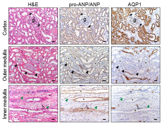
Figure 2. ANP protein expression in renal segments.
Human pro-ANP/ANP protein in the renal cortex and medullary sections was stained by immunohistochemistry. AQP1 staining was included as a control. Black arrows indicate distal convoluted tubules near glomeruli (g) (top panels). Asterisks indicate collecting ducts (middle and lower panels). Black arrowheads indicate thick ascending limbs (middle panels). Open arrowheads indicate descending thin limbs. Green arrowheads indicate ascending thin limbs. Scale bars: 40 μm.

