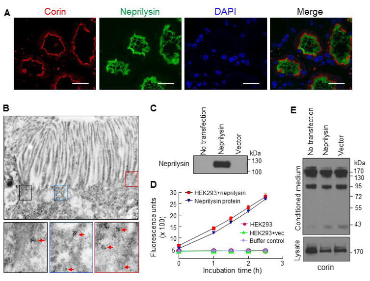
Figure 6. Corin and neprilysin expression in proximal tubules and transfected HEK293 cells.
(A) Human renal cortex sections were co-stained for corin (red), neprilysin (green), and nuclei (DAPI). Scale bar: 20 μm. (B) Apical corin expression in proximal tubular epithelial cells of mouse kidneys was verified by EM. Red arrows indicate antibody-conjugated gold particles. Original power magnifications of the top panel and lower panels were x34003 and x91568, respectively. (C and D) Plasmid expressing human neprilysin or a control vector was transfected in HEK293 cells. Neprilysin protein in cell lysates from the transfected cells was examined by Western analysis (C). Neprilysin activity in the transfected cells was verified by a fluogenic assay (D). Additional controls in the neprilysin activity assay included parental HEK293 cell lysate (no transfection), buffer (negative control), and purified neprilysin protein. (E) Corin protein fragments in the conditioned medium (top panel) and cell lysates (lower panel) from the transfected HEK293 cells were analyzed by Western blotting.

