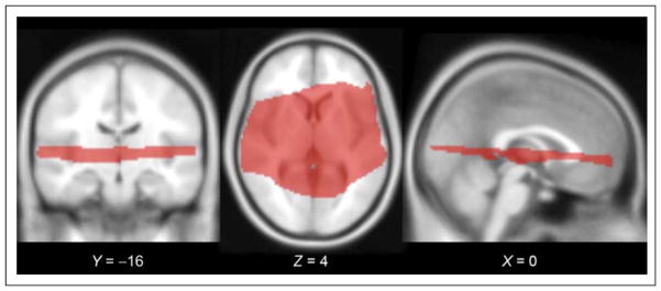
Figure 2.
Slice orientation for data collection. To maximize spatial resolution in a minimal period (TR = 3000 msec, acquisition time = 1000 msec, voxel size = 2.0 × 2.0 × 1.9 mm), we collected EPI volumes in a restricted slice set surrounding the sylvian fissure. Group-level statistical inference was restricted to the subset of voxels for which all subjects’ collected slices overlapped when transformed to standard space; this subset is shown here on the SPM MNI152 template, with MNI coordinates listed for each slice.

