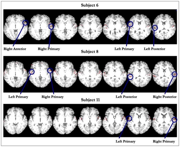
Figure 5.
Example clusters of interest from three subjects used as inputs for pattern classification. Clusters were defined based on all conditions versus rest, at a liberal threshold of p < .001 (uncorrected) with an extent threshold of 20 voxels. For each subject, we first identified the cluster of greatest spatial extent, generally in and around HG in each hemisphere. We then subdivided the STG into HG, aSTG, and pSTG regions and identified peaks of activity that fell within these anatomically defined sectors in both the left and right hemispheres. A majority of subjects had peaks in the left and right HG, with fewer having peaks in pSTG, and with the least number of peaks occurring in aSTG.

