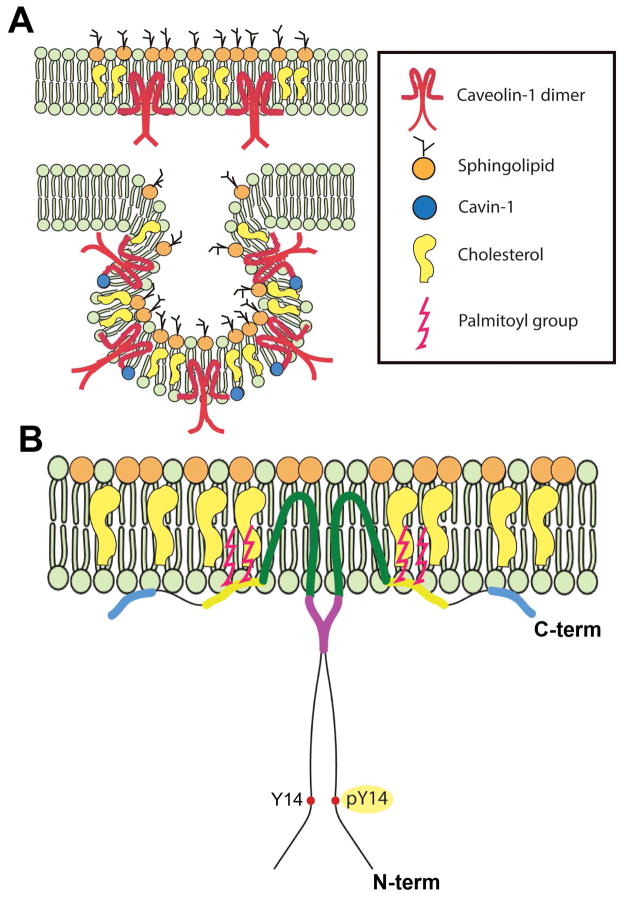
Fig. 1.
Caveolae membrane domains and the signature Cav-1 protein. (A) Illustrations of a planar Cav-1-enriched membrane microdomains with “extracaveolar” caveolin-1 and a typical caveola. (B) Model of the insertion of a caveolin-1 homodimer with the CSD (purple) and the Tyr-14 which can be phosphorylated by several tyrosine kinases.

