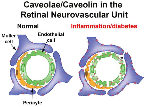
Fig. 10.
Illustration of Cav-1/caveolae in the neurovascular unit of normal and inflamed retinal vasculature. Under normal conditions, caveolae show a predominant abluminal localization in vascular endothelium (green) and mural cells (orange). Cav-1 protein, but no caveolae are detectable in Müller glia (purple). During inflammatory conditions (e.g., diabetic retinopathy), caveolae increase in number and show bipolar localization in both endothelial and mural cells possibly promoting transcellular permeability. Cav-1 expression in Müller glia is dramatically increased.

