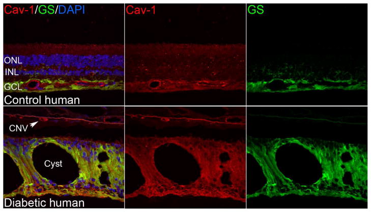
Fig. 3.
Localization of caveolin-1 and caveolae in control and diabetic human retinae. Like the murine retina, Cav-1 immunoreactivity is detectable in retinal vasculature and Müller glia (labeled with glutamine synthetase, “GS”, in green). In the cystic, diabetic retina in the lower panels Cav-1 immunoreactivity is enhanced in Müller glia and is also present in the choroidal neovascular (CNV) lesion. (Elliott laboratory, unpublished images).

