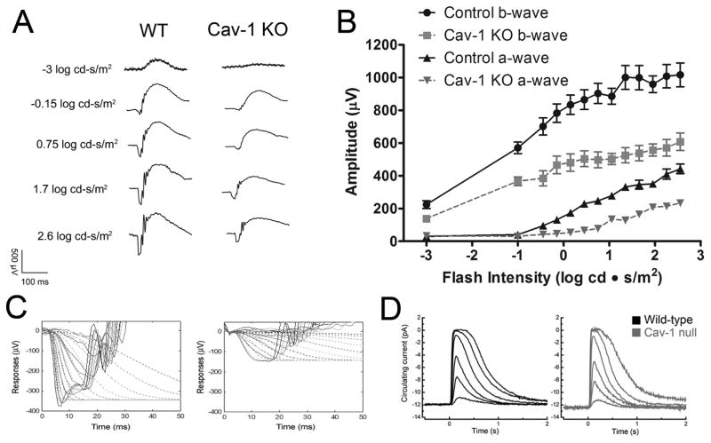
Fig. 6.
Electrophysiological studies from Cav-1−/− mice. ERG analysis (A–C) revealed significantly reduced photoreceptor a-wave and second order neuronal (b-wave) responses, in vivo. (D) Suction electrode recordings of rod light responses to graded series of flash intensities reveal normal photoresponses from isolated rods. (left panel) Mean current traces in wildtype rods (black traces) to flashes at intensities of 4, 17, 43, 160, 450, and 1122 photons μm−2. The average dark current in wildtype rods was 14.1 ± 0.6 pA (n = 45). (right panel) mean current traces from Cav-1−/− to the same flash intensities. The average dark current in Cav-1 null rods was 12.5 ± 1.1pA(n = 9), not significantly different from wildtype (p = 0.24, Student’s t-test). The flash sensitivity of the Cav-1−/− rods was 0.26 ± 0.03 pA/photon/μm2, not significantly different (p = 0.20) from wildtype rods (0.31 ± 0.02 pA/photon/μm2. These results indicate that retinal function in situ is defective but that the functional deficit is not intrinsic to rods. Reproduced from (Li et al., 2012).

