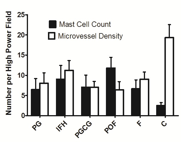
Figure 1.

Tryptase-positive mast cells in: a) control group, b) fibroma, c) inflammatory fibrous hyperplasia, d) peripheral ossifying fibroma, e) pyogenic granuloma, f) peripheral giant cell granuloma (IHC staining, ×400).

Tryptase-positive mast cells in: a) control group, b) fibroma, c) inflammatory fibrous hyperplasia, d) peripheral ossifying fibroma, e) pyogenic granuloma, f) peripheral giant cell granuloma (IHC staining, ×400).