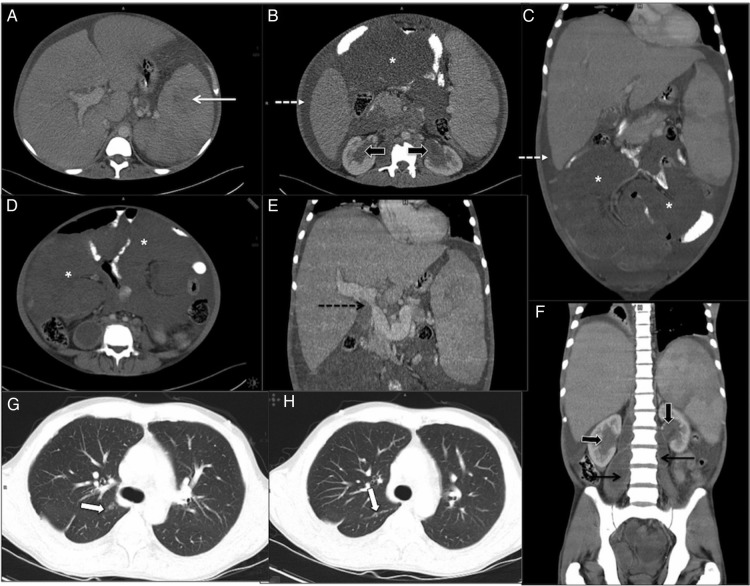
Figure 1.
(A–H): Contrast-enhanced CT in 35-year-old man with idiopathic myelofibrosis (IMF). There was hepatosplenomegaly with focal hypodense lesions in spleen (white arrow, A), multiple mesenteric masses (*, (D)), soft tissue thickening around both renal pelvis (block arrows, (B and F)) and ill-defined lung nodules in right upper lobe and abutting major fissure (white block arrows (G and H)), which were all considered either foci of extramedullary hematopoeisis (EMH) or lymphoma at initial presentation. There was also bilateral psoas abscess (black arrow, (F)) with necrotic paraortic nodes (not shown) suggestive of tuberculosis. Splenoportal axis was dilated with focal narrowing of portal vein at porta (black dotted arrow, (E)) suggestive of portal hypertension and ascites (white dotted arrow, (B and C)).

