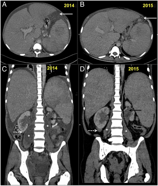
Figure 4.

(A–D): Follow-up CT scan abdomen in 2015 and comparison with 2014 scans. (A and B) showing serial axial sections for comparison. There is partial resolution of ascites with residual peritoneal nodularity (white arrows (A and B)). (C and D) showing serial coronal sections for comparison. There is resolution of ascites and bilateral psoas abscesses (*). The right infrarenal (white dashed arrow) and left parapelvic soft tissue (back arrow) increased on follow-up while spleen size was unchanged.
