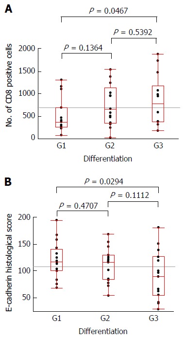
Figure 5.

The box-and-whisker plots regarding pathologically assessed differentiation and the number of CD8+ cells (A) and the E-cadherin histological score (B). Significant differences were observed in the comparison of G1 and G3 cases according to the number of CD8+ cells (P = 0.0467) and according to the E-cadherin histological score (P = 0.0294), respectively.
